
LESSON 09 · Body structure and function
The Body as a Whole: Where Organs Are and How They Connect
Understanding a body diagram means knowing where organs are, what lies beside them, and how substances and signals move between them. We establish a spatial map, then follow one breath to connect the structures into a working whole.
What you will be able to do
- Describe organ locations using the person’s own left and right and common anatomical directions.
- Distinguish the thorax, abdomen, and pelvis and locate major organs.
- Explain cooperation between systems during a breath and after a meal.
In this lesson
Establish a shared set of coordinatesHow structures fit within the chestThe abdomen and pelvis form a connected regionSystems are a way to study connected functionsFollow one breath across several systemsBuild a functional map alongside the anatomical oneBilingual termsSourcesEstablish a shared set of coordinates
Left and right belong to the person being illustrated. In a front-facing drawing, that person's right usually appears on your left. The standard anatomical position is upright and facing forward, with the arms beside the body and palms forward. Descriptions retain this reference even when the person is lying down, allowing diagrams and clinical reports to use consistent directions.
Anterior and posterior describe relative positions: the sternum is anterior to the heart. Medial and lateral refer to distance from the midline. Proximal and distal commonly describe locations nearer to or farther from a limb's attachment. Planes matter too: sagittal sections separate left from right, frontal sections separate front from back, and transverse sections separate upper from lower parts. A front view and a cross-sectional image show different perspectives. Check orientation markers before identifying structures.
Directional terms work across body regions. The elbow is nearer the upper limb’s attachment to the trunk than the wrist, so it is proximal to the wrist. That relationship remains the same whether the hand is raised or lowered, avoiding the ambiguity of everyday descriptions such as above and below.
How structures fit within the chest
The ribs, sternum, and thoracic vertebrae help protect the chest and participate in breathing movements. The lungs occupy the two sides of the thorax. The heart lies between them in the mediastinum, behind the sternum, with much of it to the left of the midline; it is not wholly confined to the left chest. The trachea divides into the main bronchi, while the esophagus passes behind it toward the stomach.
The diaphragm forms an important boundary between the chest and abdomen and is a major breathing muscle. Openings allow the esophagus and large vessels to cross this boundary. Separation therefore does not mean isolation. Find the diaphragm first when orienting a body diagram. Remember that real organs have depth, overlapping relationships, and movement during breathing; an orderly front-view illustration cannot show all of this at once.
Locate the organs, then connect their functions
Organs of the chest and abdomen
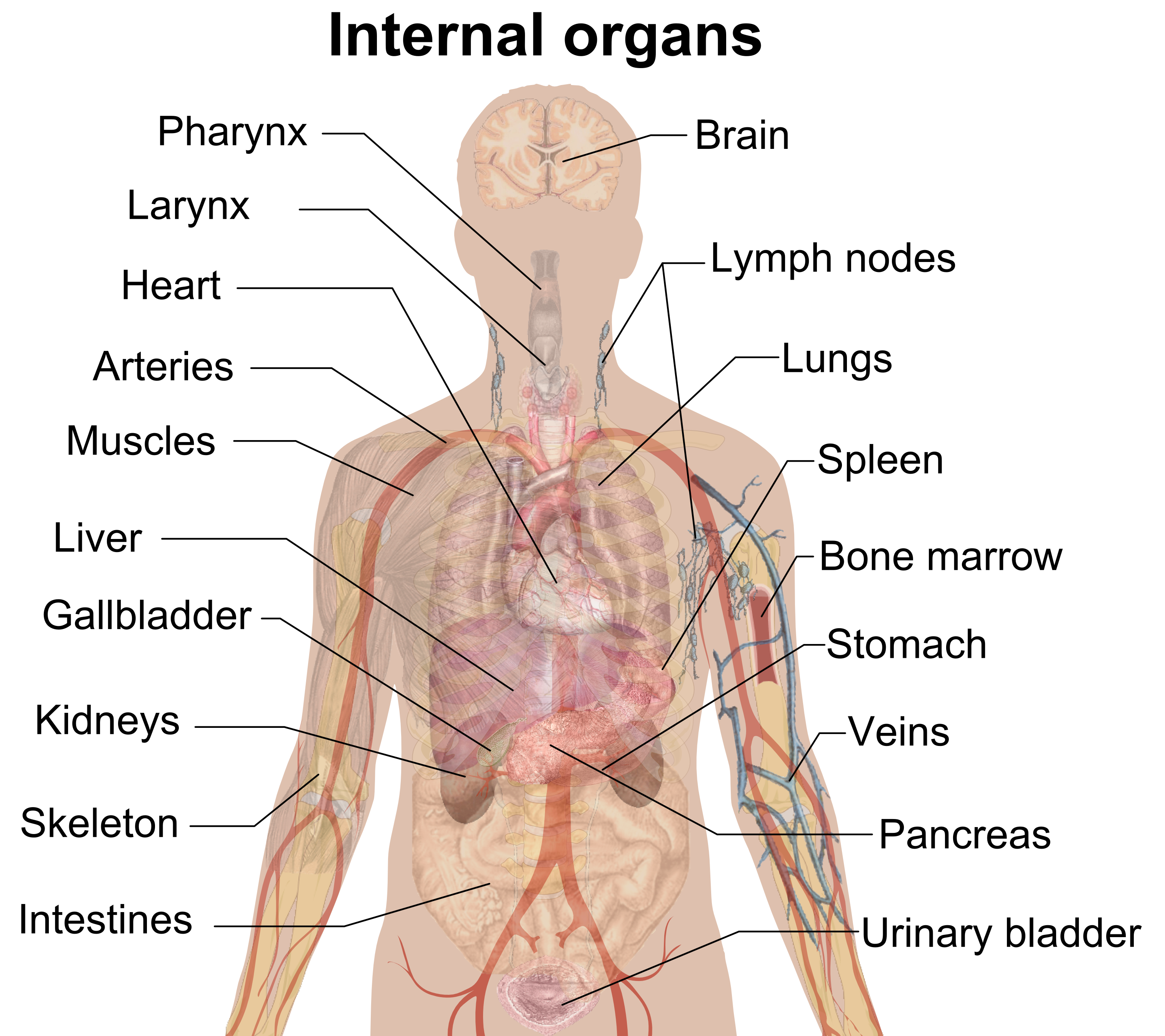
Internal organs · Mikael Häggström · CC0 / Public domain

| English label | Chinese equivalent |
|---|---|
| Heart | 心脏 |
| Lung | 肺 |
| Liver | 肝脏 |
| Stomach | 胃 |
| Kidney | 肾脏 |
Digestive anatomy
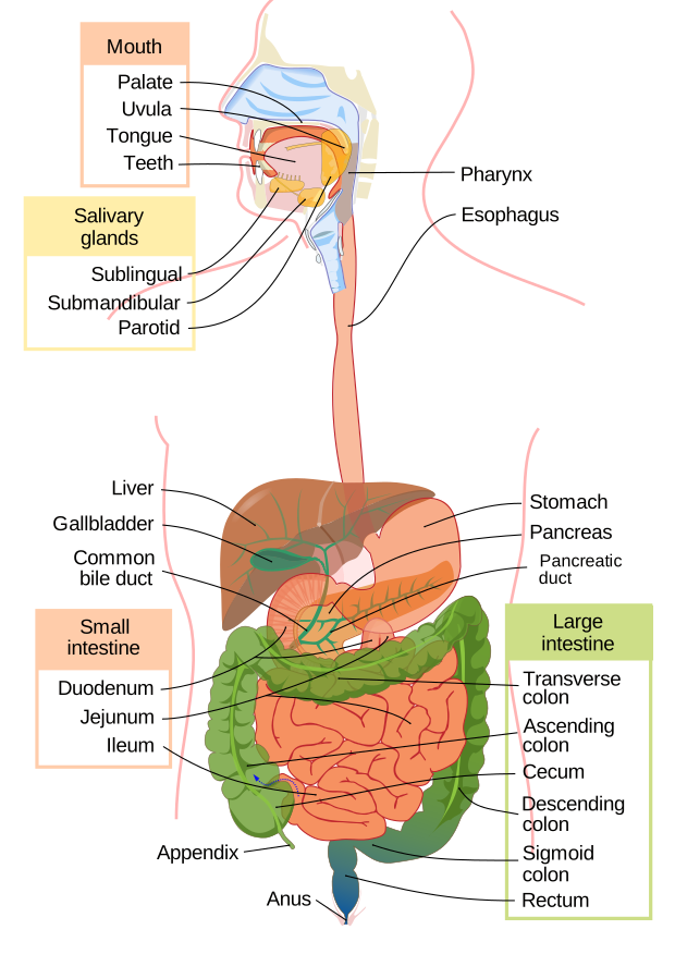
Digestive system diagram en.svg · Mariana Ruiz, Jmarchn · Public domain
Original diagrams retained; the animation was converted from GIF to MP4.


| Label in the diagram | Chinese equivalent |
|---|---|
| Liver | 肝脏 |
| Stomach | 胃 |
| Pancreas | 胰腺 |
| Small intestine | 小肠 |
| Large intestine | 大肠 |
This figure shows only the digestive system. Which structures carry food, and which contribute secretions? Then look in the lesson for links to circulation, nerves and hormones.
The abdomen and pelvis form a connected region
The liver is mainly in the upper right abdomen, the stomach mainly in the upper left, and the spleen lies farther to the upper left beside the stomach. Much of the pancreas lies behind the stomach. Small-intestinal loops occupy the abdomen, with the large intestine framing part of their outer extent. The kidneys lie near the posterior abdominal wall, behind the peritoneum, rather than hanging among the bowel loops. Diagrams often remove frontmost structures to reveal these deeper organs.
No complete muscular partition like the diaphragm separates abdomen from pelvis. The bladder, rectum, and parts of the reproductive organs occupy the pelvis, with relationships affected by anatomy and filling. These locations orient us but cannot diagnose pain. Also distinguish the digestive tube's lumen from its wall: food travels within the lumen, while absorbed substances cross an epithelial barrier into the internal environment.
Systems are a way to study connected functions
An organ usually contains several tissue types and can contribute to more than one system. The pancreas releases digestive secretions into the intestine and metabolic hormones into the blood. Kidneys form urine while also contributing to blood pressure, acid-base regulation, and red blood cell production. A single label per organ would hide these connections.
Teaching systems commonly cover circulation, breathing, digestion, urine formation, neural control, hormones, immune and lymphatic functions, movement, reproduction, and the skin. Some courses separate muscle and skeleton; others teach them together. Immune and lymphatic functions may also be organized differently. The count therefore varies with the classification used; the functions and connections are what matter for learning. A more useful set of questions is: what enters, what changes, what leaves, and which other structures supply fluid, blood, or signals?
Follow one breath across several systems
During quiet inhalation, neural signals activate the diaphragm and other breathing muscles. The chest expands, the pleural mechanical connection helps the lungs expand with it, and pressure inside the lungs briefly falls below outside pressure, drawing air through the airways. One pleural layer covers the lungs and another lines the chest wall. A thin film of fluid between them helps the lungs move with the chest wall. Air normally enters the airways and alveoli, not the pleural space. In the alveoli, a very thin barrier separates air from blood in pulmonary capillaries.
Oxygen crosses down its partial-pressure gradient and is carried mainly by hemoglobin in red blood cells. Blood returns through pulmonary veins to the left heart and is pumped to the systemic circulation. Carbon dioxide produced by tissues travels back in blood, crosses into alveoli, and is exhaled. The sequence requires neural control, muscle movement, open airways, an exchange barrier, and circulation. Ventilation, gas exchange, and delivery to tissues are connected but distinct processes.
Build a functional map alongside the anatomical one
A meal offers another example. The digestive tract breaks down food and the small intestine absorbs nutrients. Glucose and amino acids travel first through the portal circulation to the liver, while most absorbed long-chain fats enter intestinal lymph before reaching blood. Pancreatic hormones help regulate nutrient use and storage. Food itself does not pass through the liver or pancreas; participating in digestion differs from being part of the food's route.
Keep two simple drawings as you study each system: one shows organ locations, and the other shows where materials and signals go. Use different line styles for air, food, blood, and neural signals, and label what each arrow represents. If an arrow jumps from an alveolus straight to leg muscle, look for the missing blood and heart connections. Explaining those intermediate steps turns a list of organs into an account of how the body works.
The two maps check each other. One shows proximity; the other shows actual connections. Nearby structures need not open into one another, while distant organs may cooperate through vessels or nerves. These questions provide a useful framework for the detailed heart, lung, and digestive lessons that follow.
Apply what you have learned
Draw two simple diagrams. First locate the diaphragm, heart, lungs, liver, stomach, and kidneys. Then trace the major steps by which oxygen in air reaches leg muscle. Why does the second drawing need more than lungs and a leg?
Read the explanation
Label the individual’s own left and right and represent the kidneys as posterior structures. The functional drawing should include airways, alveoli and the exchange barrier, pulmonary capillary blood, pulmonary veins, the left heart, and systemic vessels leading to tissue capillaries. Circulation transports oxygen after it enters blood; oxygen then crosses into tissue. Neural signals and breathing muscles enable ventilation. Arrows trace oxygen movement, not red blood cells leaving vessels to enter muscle.
Bilingual terms
- 解剖学姿势 · Anatomical position
- A standard reference posture: upright, facing forward, arms at the sides, and palms facing forward.
- 纵隔 · Mediastinum
- The central region of the thorax between the lungs, containing the heart and other structures.
- 管腔 · Lumen
- The inside space of a tubular structure, such as the intestinal space through which food passes.
Sources and further reading
- OpenStax:解剖学术语
- OpenStax:人体结构的组织层次
- OpenStax:解剖学与生理学概览
- NHLBI:呼吸系统的结构
- NHLBI:吸气与呼气
- NHLBI:呼吸调节
- NHLBI:心脏中的血流
- NIDDK:消化系统怎样工作
- NIDDK:肾脏怎样工作
- OpenStax:化学消化与吸收
Original course source-check record: 9 September 2026. Full Chinese and English sentence-by-sentence language review: 14 September 2026. AI editing and language review are not human clinical review. Linked institutions have not participated in or endorsed this course.
A moment in nature

Puffin Flying by Flowers.jpg · Eric Kilby · CC BY-SA 2.0
Converted to WebP; thumbnails may be cropped.
