
LESSON 10 · Body structure and function
How the heart beats
Place your fingertips lightly on your wrist and you can feel a pulse. Its rhythm alone does not tell you how much blood the heart ejects with each beat, or show you the valves opening and closing. To understand a heartbeat, we need to connect anatomy, electrical activity, muscle contraction and blood flow.
What you will be able to do
- Explain blood flow and valve position using pressure differences.
- Connect the cardiac cycle to electrical activation and contraction.
- Calculate cardiac output and recognise the limits of an isolated number.
In this lesson
Four chambers and two connected circuitsWhy valves open and closeA heartbeat in slow motionFrom electrical activation to contractionDoes a faster heart always pump more blood?The heart muscle needs its own blood supplyCheck your understandingBilingual termsSourcesFour chambers and two connected circuits
The heart has a right atrium, right ventricle, left atrium and left ventricle. Atria receive returning blood; ventricles pump it into arteries. Left and right refer to the person's body. In a front-facing diagram, the person's right usually appears on your left.
Blood returning from the body enters the right atrium and passes to the right ventricle, which pumps it to the lungs. In the pulmonary capillaries, blood releases carbon dioxide and takes up oxygen. It returns through the pulmonary veins to the left atrium. This is the pulmonary circuit. The left ventricle then sends blood through the aorta and its branches to the body; veins return it to the right atrium. This is the systemic circuit. The two circuits are connected in series.
Oxygen exchange takes place between tissue capillaries and surrounding cells; red blood cells normally remain within blood vessels. Blue in a diagram denotes lower oxygen content, not the actual colour of blood.
This route describes where blood goes. It does not mean the right ventricle finishes its work before the left begins. Both ventricles contract at approximately the same time. Heart anatomy and circulation
Why valves open and close
The tricuspid valve lies between the right atrium and ventricle; the mitral valve lies between the left atrium and ventricle. The pulmonary and aortic valves guard the ventricular outlets. The tricuspid and mitral valves are collectively called the atrioventricular valves.
Valve movement depends on pressure differences. Contraction changes pressure within a chamber. A forward pressure difference opens the appropriate valve; reversal of that difference tends to close it, limiting backflow. Valves do not actively pump blood.
When left ventricular pressure exceeds left atrial pressure, the mitral valve closes. Pressure must rise further before the aortic valve opens. The chordae tendineae and papillary muscles support the atrioventricular valves during contraction, preventing the leaflets from prolapsing into the atria. They do not pull the valves open. Valve locations; valve support structures
Examine a heartbeat in stages
Heart anatomy
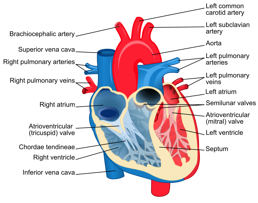
Heart diagram-en.svg · ZooFari · CC BY-SA 3.0
Original diagrams retained; the animation was converted from GIF to MP4.

Watch a heartbeat
CG Heart.gif · DrJanaOfficial · CC BY-SA 4.0
Original diagrams retained; the animation was converted from GIF to MP4.

The displayed pressures are invented snapshots in mmHg, ignoring flow inertia and valve-transition details. The phases are not equal in duration. Both ventricles work approximately together.
A heartbeat in slow motion
The cardiac cycle is a complete sequence of contraction and relaxation. We can follow four phases in the left ventricle. The right side undergoes similar changes at different pressures; valve events on the two sides are not exactly simultaneous.
Filling. Ventricular pressure is below atrial pressure. The mitral valve is open and the aortic valve closed. Blood flows into the ventricle; atrial contraction adds to filling near its end.
Isovolumetric contraction. Contraction begins. The mitral valve closes while the aortic valve remains closed. Pressure rises, but ventricular blood volume stays nearly constant. “Isovolumetric” refers to volume, not an absence of muscle activity.
Ejection. Ventricular pressure becomes sufficient to open the aortic valve. Blood leaves the ventricle, reducing its volume. The ventricle does not empty completely.
Isovolumetric relaxation. The aortic valve closes; the mitral valve has not yet opened. Pressure falls while blood volume remains nearly constant. Once ventricular pressure falls below atrial pressure, filling resumes.
Contraction therefore includes a period without ejection, and relaxation starts before filling. Watch pressure, valves and volume together. Cardiac cycle
From electrical activation to contraction
A normal heartbeat usually begins in the sinoatrial node in the right atrium. Its pacemaker cells generate rhythmic electrical activity. Activation spreads through the atria, slows at the atrioventricular node, and then travels through specialised conducting tissue to the ventricles. This sequence coordinates atrial and ventricular contraction. Autonomic nerves and hormones modify heart rate and other aspects of cardiac activity. Electrical conduction
In ventricular muscle cells, electrical activation opens calcium channels. Calcium entering the cell triggers additional calcium release from internal stores. The rise in calcium allows contractile proteins to interact and use energy from ATP to generate force. Relaxation requires calcium to be returned to storage or removed from the cell.
This connection between electrical activation and contraction is called excitation–contraction coupling. An electrocardiogram records cardiac electrical activity detected at the body surface. It does not directly measure the volume of blood ejected. Understanding pumping function also requires information about structure and mechanical activity. Cardiac muscle and electrical activity
Does a faster heart always pump more blood?
Stroke volume is the amount ejected by one ventricle per beat. Cardiac output is the amount it ejects per minute:
Cardiac output = heart rate × stroke volume.
In an invented teaching example, 70 beats per minute multiplied by 70 millilitres per beat gives 4,900 millilitres, or 4.9 litres, per minute. These values illustrate a calculation; they are not a standard for judging an individual's health.
Heart rate alone leaves stroke volume unknown. Filling, contractile ability and the load against which the ventricle ejects all influence stroke volume. A very fast rate also shortens the time available for filling. Faster does not automatically mean more effective pumping. In a steady state, the two ventricles have matching average outputs over time; adding them would wrongly count the same circulation twice. Cardiac output and its regulation
The heart muscle needs its own blood supply
Blood constantly passes through the chambers, but the myocardium obtains its oxygen and nutrients mainly through the coronary arteries and their branches. These arise at the aortic root and supply capillaries within the muscle. Venous blood then drains back towards the heart.
Blood passing through a chamber and adequate blood reaching the heart muscle are distinct matters. This distinction prepares us to understand how coronary artery disease can affect the heart itself. Coronary blood supply
Check your understanding
1. The left ventricle is contracting, but the aortic valve is closed. Is this contradictory?
No. During isovolumetric contraction, pressure is rising but has not yet reached the condition required for the aortic valve to open. Compare ventricular pressure with aortic pressure to decide whether ejection can begin.
2. Someone has the same heart rate as the example. Can you calculate their cardiac output?
Not without their stroke volume. Substituting the example's 70 millilitres would give a numerical answer based on an unsupported assumption.
3. Does observing electrical activity prove that coronary supply and pumping function are normal?
No. Electrical activity, myocardial blood supply and the mechanical effects of contraction are related, but they are not equivalent. State what the observation establishes and what remains unknown.
Apply what you have learned
In a simplified model, left atrial, left ventricular and aortic pressures are 8, 40 and 80 mmHg. Assume normal valves and ignore flow inertia. What are the valve positions? Can you distinguish contraction from relaxation from this snapshot?
Read the explanation
Both valves are closed. Ventricular pressure is higher than atrial pressure and lower than aortic pressure. The snapshot alone cannot distinguish isovolumetric contraction from relaxation; the pressure trend and surrounding phases are needed. These are invented teaching values.
Bilingual terms
- 心房 · Atrium
- A chamber receiving returning blood.
- 心室 · Ventricle
- A chamber pumping blood into an artery.
- 二尖瓣 · Mitral valve
- The valve between the left atrium and ventricle.
- 每搏量 · Stroke volume
- The volume ejected by one ventricle per beat.
- 心输出量 · Cardiac output
- The volume pumped by one ventricle per minute.
- 心肌 · Myocardium
- The muscle tissue of the heart wall.
Sources and further reading
- NHLBI: How Blood Flows through the Heart
- NHLBI: How the Heart Beats
- OpenStax: Heart anatomy
- OpenStax: Cardiac muscle and electrical activity
- OpenStax: Cardiac cycle
- OpenStax: Cardiac physiology
Original course source-check record: 9 September 2026. Full Chinese and English sentence-by-sentence language review: 14 September 2026. AI editing and language review are not human clinical review. Linked institutions have not participated in or endorsed this course.
A moment in nature

Passiflora caerulea (makro close-up).jpg · Petar Milošević · CC BY-SA 4.0
Converted to WebP; thumbnails may be cropped.
.jpg)